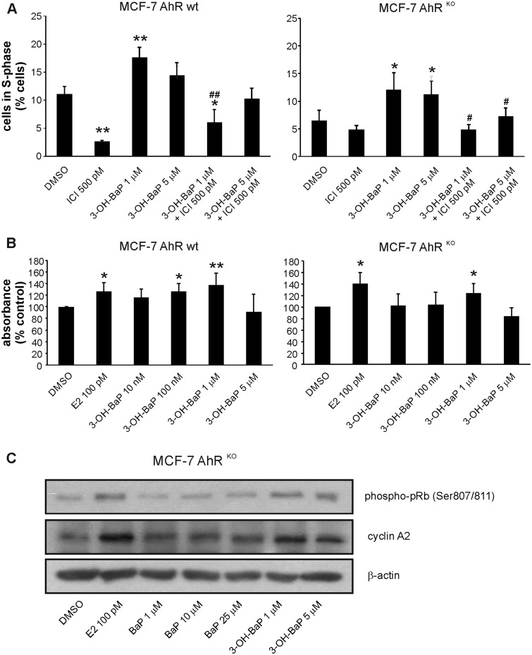
Figure 5.
The major BaP metabolite identified in MCF-7 cells, 3-OH-BaP, induces estrogen-like effects on cell cycle progression and proliferation in MCF-7 AhRKO cells. A, MCF-7 AhR wt and MCF-7 AhRKO cells were synchronized in experimental medium (phenol red-free DMEM/F12, supplemented with 5% dextran/charcoal-treated FBS) for 48 h and then exposed to DMSO (0.1%; negative control) or indicated concentrations of 3-OH-BaP, alone, or in combination with synthetic ER antagonist, ICI 182,780 (ICI; 500 pM), for 24 h. Percentage of cells in S-phase was determined using flow cytometry as described in Materials and Methods. The results represent means ± SD of three independent experiments. Symbol “*” denotes significant difference (p < .05), as compared with DMSO-treated cells. Symbol “**” denotes significant difference (p < .01), as compared with DMSO-treated cells. Symbol “#” denotes significant difference (p < .05) between the respective 3-OH-BaP treatment group and its combination with ICI. Symbol “##” denotes significant difference (p < .01) between the respective 3-OH-BaP treatment group and its combination with ICI. B, MCF-7 AhR wt and MCF-7 AhRKO cells were synchronized in experimental medium for 48 h and then exposed to DMSO (0.1%; negative control), E2 (100 pM; positive control), or indicated concentrations of 3-OH-BaP for 120 h. Cell proliferation was estimated using WST-1 assay as described in Materials and Methods. The results of absorbance detection were expressed relative to negative control (DMSO) and they represent means ± SD of three independent experiments. Symbol “*” denotes significant difference (p < .05), as compared with DMSO-treated cells. Symbol “**” denotes significant difference (p < .01), as compared with DMSO-treated cells. C, MCF-7 AhRKO cells were synchronized in experimental medium for 48 h and then exposed to DMSO (0.1%; negative control), E2 (100 pM; positive control), or indicated concentrations of BaP and 3-OH-BaP for 24 h. Cells were collected, lysed, and protein levels of cyclin A2 and phosphorylated pRb (Ser 807/811) were determined by Western blotting. β-Actin was used as a loading control. The results shown here are representative of three independent experiments.

