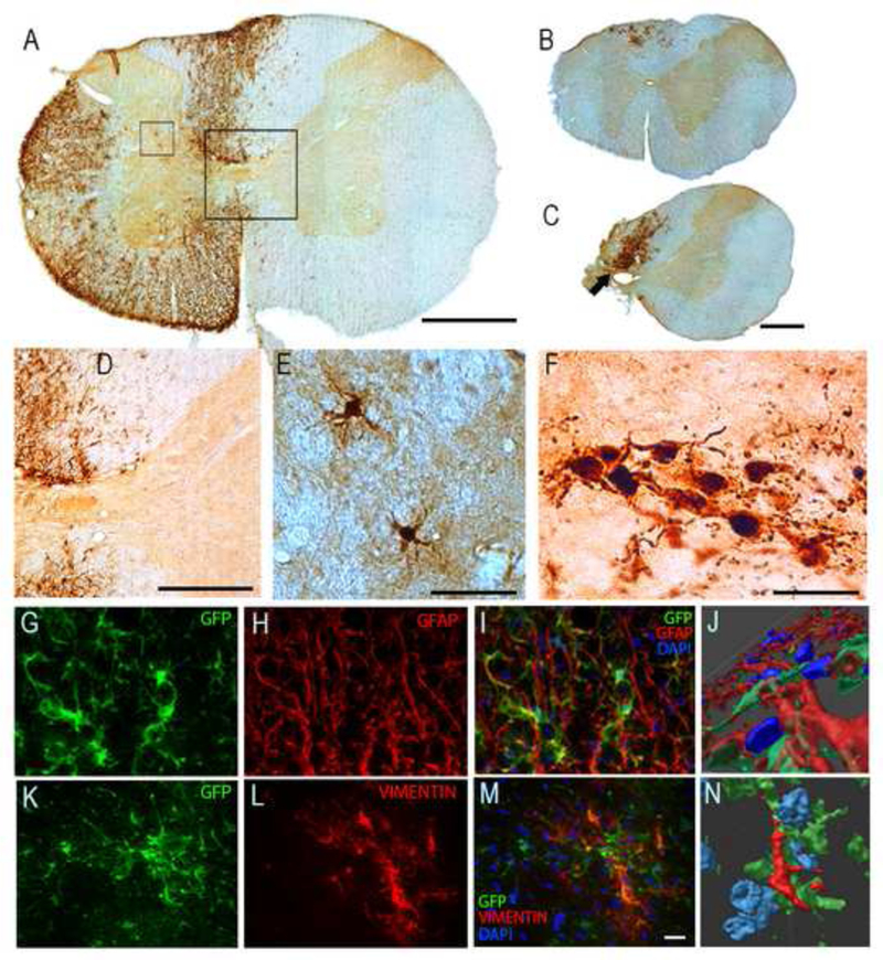
Fig. 2. Migration and differentiation of SVZ-derived NPCs after transplantation into the injured cervical spinal cord.

Panel A shows a C3 transverse histological section at the approximate site of the NPC injection (i.e., immediately caudal to the C2 injury) obtained 8-wks following the transplantation procedure. Panel B shows a section from the C1/C2 border which illustrates the rostral migration of transplanted cells. A histological example obtained at the site of the C2Hx injury is shown in Panel C. Panels D and E show higher magnification views of the areas highlighted by the boxes in Panel A. The image shown in panel F is a magnified view of the region indicated by the arrow in Panel C. Panels G-N show histological examples which demonstrate co-labeling of the transplanted cells with GFAP (marker for astrocytes) and vimentin (a cytoskeletal intermediate filament protein). In the sequence of images shown in G- I, the same section of cervical white matter is shown to illustrate co-localization of the transplanted SVZ-derived neurons (which are GFP-positive and therefore green) with GFAP (red) and DAPI (blue). The same area of tissue is shown as a three dimensional rendering in Panel J. These images confirm that a subset of the transplanted NPCs developed along an astrocytic lineage. Panels K-M demonstrate co-localization of transplanted GFP-positive cells with vimentin (red) and DAPI (blue), and a three dimensional rendering is provided in N. These images are representative of the data used to determine that subsets of the transplanted neurons were co-labeled with vimentin, which is also consistent with an astrocytic phenotype. Scale bars: 500 pm in panels A-C, 200pm in panel D, 50 pm in panels E-F, and 20 pm in panels G,H,I, K, Land M.
