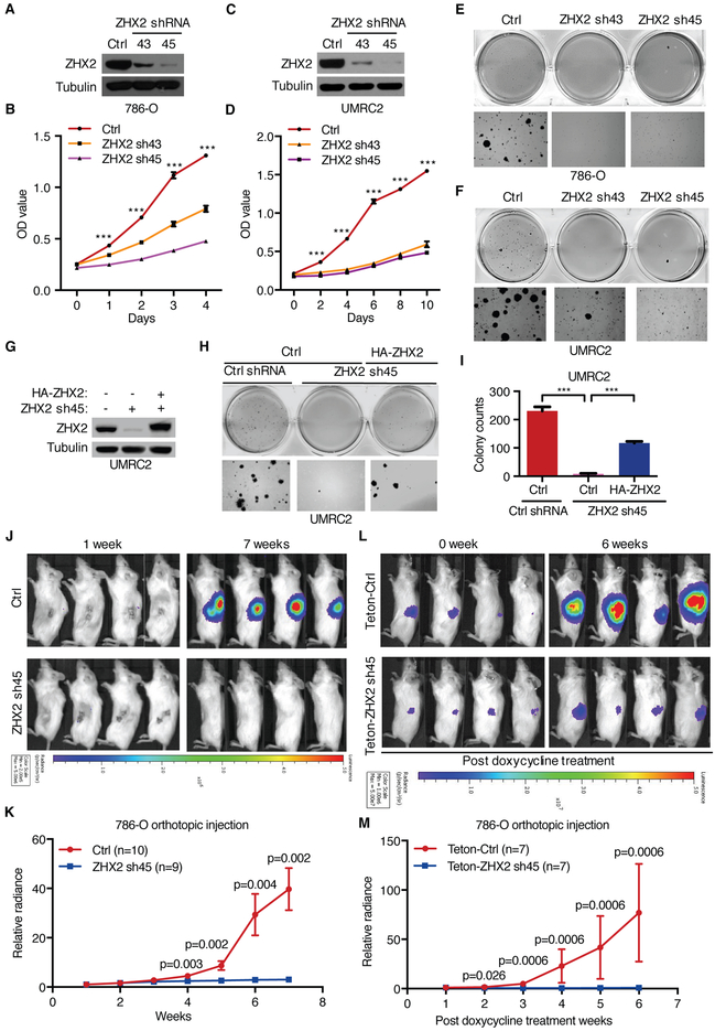
Figure 3. Requirement of ZHX2 for ccRCC cell proliferation, anchorage-independent growth and tumorigenesis.
(A-F) IB of cell lysates (A, C), cell proliferation (B, D) and soft agar growth (E, F) of 786-O and UMRC2 cells infected with lentivirus encoding control (Ctrl) or ZHX2 shRNAs (43, 45) (N=3). See fig. S4A-B for soft agar quantitation results.
(G-I) IB of cell lysates (G) and representative soft agar growth assays (H) and their quantification (I) of UMRC2 cells transfected with ZHX2 sh45-resistant HA-ZHX2 or control (Ctrl) vector, followed by ZHX2 sh45 or control (Ctrl) shRNA infection (N=3).
(J-M) Representative bioluminescence imagings of 1 and 7 weeks post-implantation (J) and quantification of bioluminescence imaging (K) from 786-O cells luciferase stable cells infected with either ZHX2 sh45 or control (Ctrl) shRNA, or imagings of 0 week and 6 weeks post-doxycycline treatment (L) and quantification of imaging (M) from 786-O luciferase stable cells infected with lentivirus encoding either Teton-ZHX2 sh45 or Teton-control (Teton-Ctrl) shRNA injected orthotopically into the renal sub-capsule of NOD scid gamma (NSG) mice as indicated. The Mann-Whitney test was used to calculate the p values.
Error bars represent SEM, ***P<0.001 (unpaired t-test) in panel B, D and I.

