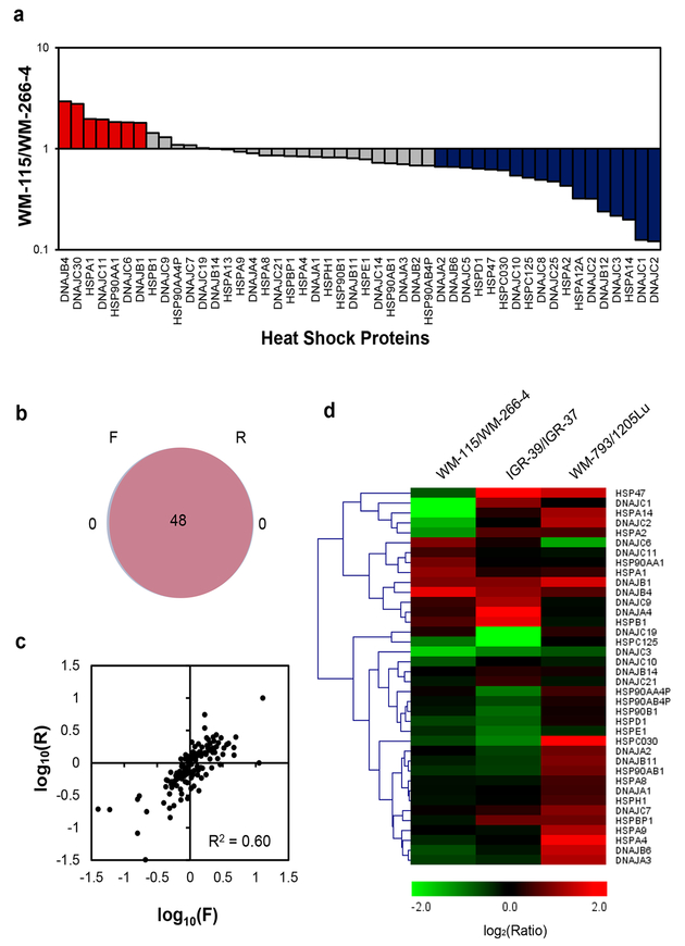
Figure 2.
Performances of PRM-based targeted proteomic approach for interrogating the perturbations in expression of heat shock proteins during melanoma metastasis. (a) Differential expression of heat shock proteins in WM-115/WM-266–4 paired melanoma cells. (b) A Venn diagram displaying the overlap between quantified heat shock proteins from the forward and reverse SILAC labelings of WM-115/WM-266–4 paired melanoma cells. (c) Correlation between the ratios obtained from forward and reverse SILAC labeling experiments. (d) A heat map showing the differences in expression of heat shock proteins in three pairs of primary/metastatic melanoma cell lines. Genes were clustered according to Euclidean distance. The data in (a) and (d) represent the mean of the results obtained from one forward and one reverse SILAC labeling, and Table S3 lists the ratios obtained from individual measurements.

