
Fig. 1.
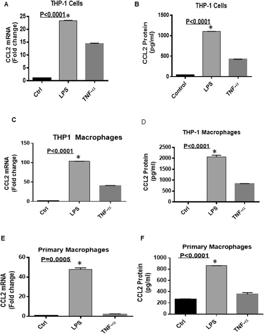
Effect of LPS on CCL2 expression in human monocytic cells and macrophages. THP-1 cells were treated with LPS (10 ng/ml), TNF-α (10 ng/ml; positive control) and PBS Control = Ctrl) for 24 h. Cells and culture media were collected. Total RNA was isolated and CCL2 mRNA was quantified by real time PCR. Relative mRNA expression was expressed as fold expression over average of gene expression in BSA treated cells. LPS significantly increased the expression of CCL2 in THP-1 (A). Secreted CCL2 protein was increased in culture media was determined by ELISA. LPS induced high production of CCL2 compared to control (B). THP-1 cells were converted into macrophages and were treated with LPS or TNF- α for 24 h. Cells and culture media were collected. Real time PCR data showed increased CCL2 mRNA expression in LPS treated macrophages compared to control (C). LPS induced CCL2 protein in culture media (D). Primary macrophages were treated with LPS orTNF- α for 24 h. Cells and culture media were collected. Real time PCR data show that LPS increased expression of CCL2 (E). Secreted CCL2 in culture media was determined by ELISA and which was significantly upregulated by LPS (F). Data are shown as mean ± SEM of three independent experiments