
Fig. 4.
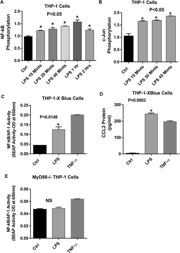
LPS activates NF-kB and AP-1 transcription factors. THP- 1 cells were treated with LPS for different time points and cell lysates were prepared as described in methods. Samples were run for Quantikine assay on PathScan Sandwich Elisa kit. Our data showed that phosphorylated NF-kB and c-Jun was seen in LPS treated cells (4A and 4B). THP-1-XBlue cells (THP-1 cells stably expressing a secreted embryonic alkaline phosphatase (SEAP) reporter inducible by NF-κB and AP-1) were treated with LPS or PBSor TNF-α for 24 h. Culture media were collected. Cell culture media were assayed for SEAP reporter activity (degree of NF-κB / AP-1 activation) along with CCL2 production. LPS increased NF-kB/AP-1 activity as compared to control (C and D). Similarly, THP- 1-XBlue™-defMyD cells (Cells deficient in MyD88 activity) were treated with LPS (10 ng/ml) or TNF-α (10 ng/ml) for 24 h. SEAP reporter activity (degree of NF-κB /AP-1 activation) was determined in the cell culture media. MyD88 deficiency inhibits the LPS induced activation of NF-kB/AP-1 (E). The results obtained from three independent experiments are shown. The data are presented as mean ± SD