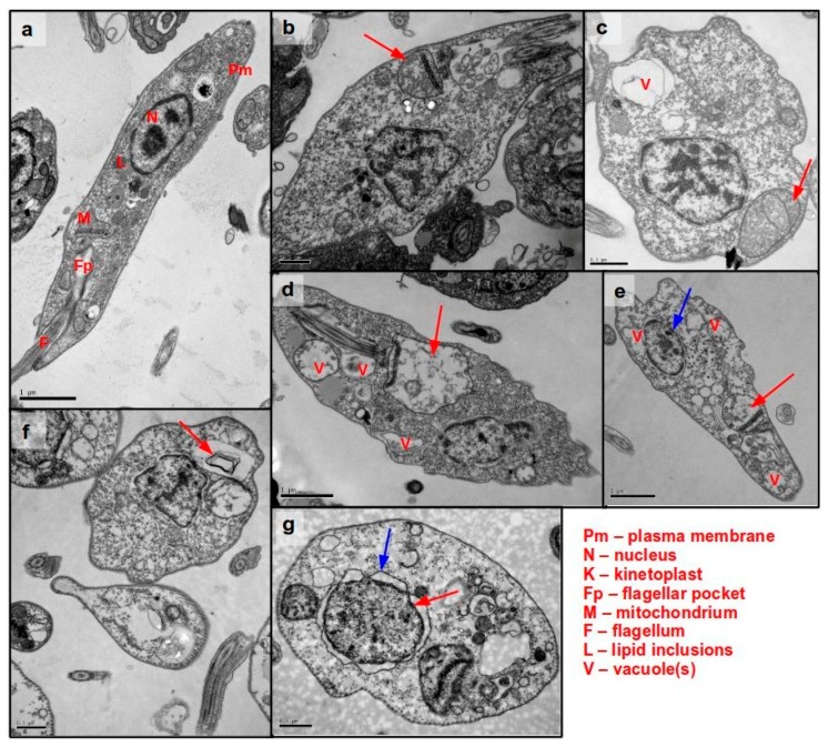
Figure 4.
Transmission electron microscopy imaging of L. (L.) infantum promastigotes treated with helenalin acetate (1). 2 × 10⁷ promastigotes/well were incubated with 1 for different periods of time. (a) Untreated control. (b) 0.5 h; (c) 1 h; (d) 2 h; (e,f) 3 h; (g) 4 h. The figure shows representative images taken from one out of two independent experiment. The observed ultrastructural observations appeared consistently in both independent Transmission Electron Microscopy (TEM) experiments.

