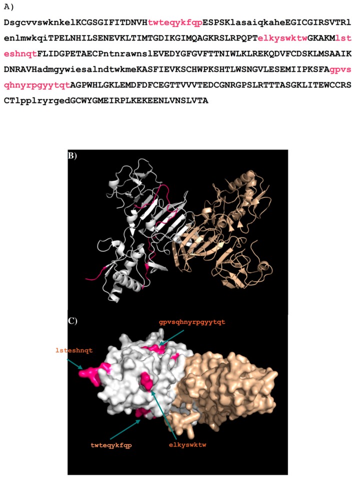
Figure 12.
(A) The predicted epitopes of DENV2 NS1 protein to antibody GUS2 are highlighted in lower case and coloured magenta in the protein. The peptides identified as binders using the sequence search only are shown in lower case. The selection criteria based on their surface exposure and secondary structure is described in detail in the supplementary section (Figure S3); (B) Backbone presentation of the dimer form of DENV2 NS1 protein showing the predicted epitopes in magenta; (C) Surface presentation of the dimer form of DENV2 NS1 protein showing the predicted epitopes in magenta. Note that the majority of the loop (residues 108–128) is missing in the crystal structure of DENV2 NS1 protein (PDB code 4O6B) so that the epitope B “elkyswktw” and epitope C “lsteshnqt” are only illustratively displayed.

