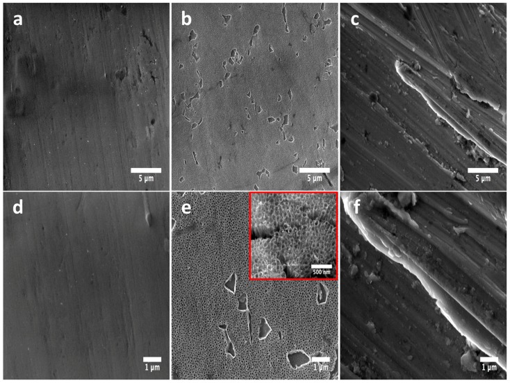
Figure 1.
FE-SEM micrographs illustrating the surface morphology of the experimental materials: (a) Flat-Ti6Al4V surface showing a flat and smooth surface; (b) anodized 80 nm nanotubes (NTs) highlighting the homogenous formation of a nanostructured layer; (c) Rough-Ti6Al4V surface presenting irregular grooves among the material; (d) high zoom of the flat surface demonstrating the non-presence of a nanostructured surface; (e) high amplification of the NTs confirming the nanotubular homogeneity (inset represents a superior magnification, which described the nanotubular morphology); (f) high magnification of the rougher surface.

