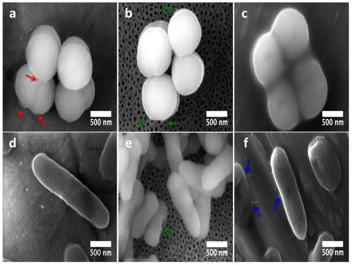
Figure 10.
Nano-scale comparative bacteria-surface interactions among Gram-positive and Gram-negative microorganisms over the surfaces at 6 h: (a) S. epidermidis on the Flat-Ti6Al4V material (the red arrows highlight the huge contact bonds among the surface and the cell-cell interactions); (b) 80 nm NTs illustrating decreased adhesion of S. epidermidis (green arrows points out tiny surface adhesion-bonds); (c) S. epidermidis adhesion on the rough material; (d) P. aeruginosa largely disseminated on the Flat Ti6Al4V; (e) deformed bacilli interaction on NTs (the green arrow represents deformed morphology of direct interacting bacteria to the NTs); (f) P. aeruginosa growth on the rough material (the blue arrows show the bacterial interaction bonds above the surface).

