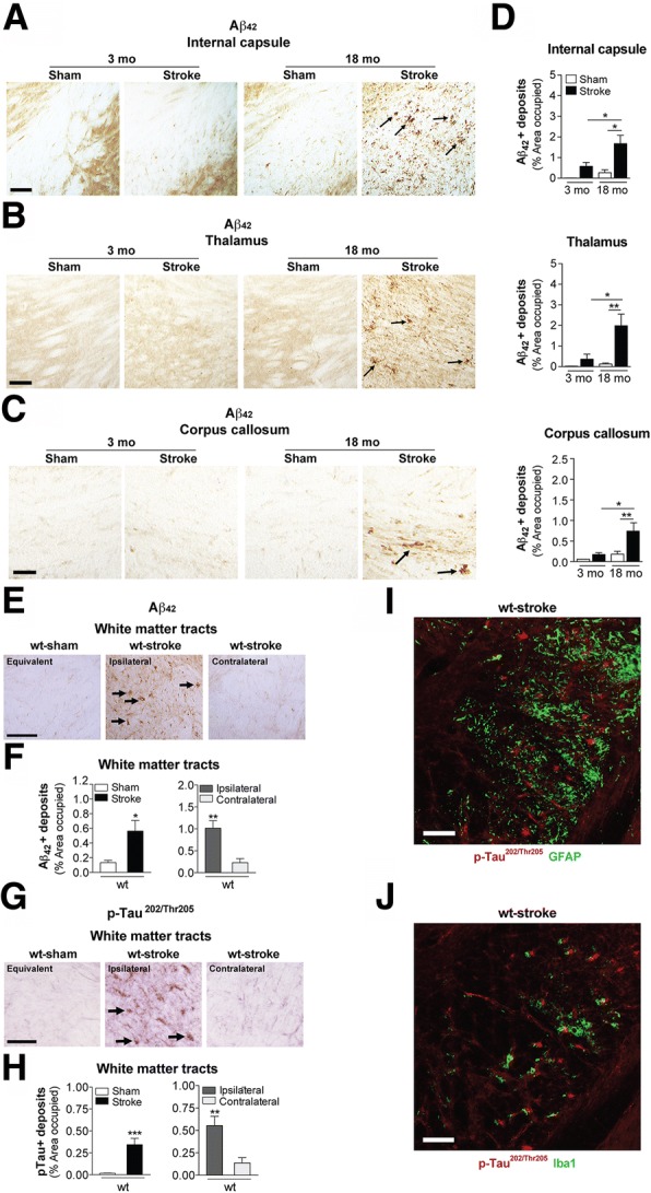
Fig. 3.

Stroke causes β-amyloid (Aβ) and phosphorylated (p) tau deposition in the white matter tracts of aged wildtype (wt) mice compared to young adult mice. Representative 10× images of Aβ42-immunolabeled deposits (arrows) in the white matter tracts of the (a) internal capsule, b thalamus, and (c) corpus callosum of the 3 and 18 mo, sham- and stroke-operated C57BL/6 mice at 8 weeks post-surgery. Scale bar, 100 μm (internal capsule and thalamus), 50 μm (corpus callosum). Nissl-stained sections to the left of each image delineate where representative images were taken. d Quantification of the ipsilateral hemisphere revealed a significant deposition of Aβ42 in the internal capsule (top graph), thalamus (middle graph), and corpus callosum (bottom graph) of the 18 mo stroked mice relative to the age-matched sham-operated mice. Furthermore, the 18 mo stroked mice had significantly more Aβ42 accumulation in three of the brain regions analyzed compared to the 3 mo stroked mice. e-h Representative 10× images of (e) Aβ42- and (g) p-tau-immunolabeled deposits (arrows) in white matter tracts (thalamus-internal capsule) of the 18 mo mice at 12 weeks after sham or stroke surgery (Equivalent = area imaged in wt-sham mice that is equivalent to the ipsilateral hemisphere imaged in wt-stroke mice; Contralateral = area imaged in the contralateral hemisphere of wt-stroke mice that is equivalent to the ipsilateral hemisphere of wt-stroke mice). Scale bar, 125 μm (Aβ42 and p-tau). Quantification of the ipsilateral and contralateral hemispheres revealed significantly more deposits of (f) Aβ42 and (h) p-tau in the white matter tracts of the 18 mo stroked mice compared to the age-matched sham-operated mice. Furthermore, there was also significantly more Aβ42 and p-tau accumulation in the white matter tracts of the ipsilateral versus the contralateral hemisphere. No Aβ42 signal was detected in (i) astrocytes (GFAP, green; n=3 mice/experimental group) or (j) microglia (Iba1, green; n=3 mice/experimental group). Scale bar, 125 μm. Data represent mean ± SEM. *p<0.05, **p<0.01, and ***p<0.001
