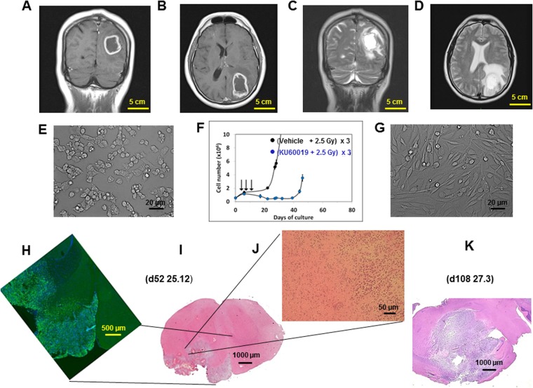
Figure 3.
The GIC-driven COMI tumor. (A,B) Patient’s tumor i: coronal and axial T1-weighted post-contrast MRI showing a left parieto-occipital intra-axial tumor with irregular contrast enhancement and necrotic core. (C,D) Patient’s tumor ii: T2-weighted images showing significant peritumoral edema and infiltrative behavior. (E) GIC i: the GIC component of the tumor was isolated immediately after surgery. Under matrigel-coating and serum-free conditions, the cells grew and layered onto a monolayer, maintaining intact self-renewal capacity. (F) GIC ii: the COMI GIC could be effectively radiosensitized in vitro by exposure to 1 µM KU60019 30 min prior to irradiation with three 2.5 Gy IR fractions [reproduced from12, with permission]. (G) GIC iii: removal of growth factors and addition of 10% FCS to the proliferation medium after approximately two weeks induced GIC differentiation with acquisition of astrocytic morphology and altered refractory index. (H–K) Orthotopic tumor. 3 × 10⁵ COMI serum-free grown GIC were stereotactically injected in the left corpus striatum of immunodeficient NOD SCID mice. Staining with hematoxylin/eosin of brain tissue sections revealed 52 (I,J) and 108 (K) days later a growth pattern of the orthotopic tumor reflecting the characteristics of the clinical tumor: a voluminous expansive lesion (I,K) with an irregular infiltrating wall (J) exerting mass effect on the adjacent structures (K). Immunostaining at d52 revealed that most of the orthotopic tumor expressed the stem cell marker nestin, indicating its stem cell-driven character (H). Mouse ID numbers 25.12 and 27.3 are indicated for the sake of reference with the days (d) of tumor development.

