
Fig. 5. Structural comparison between rhodopsin complexes.
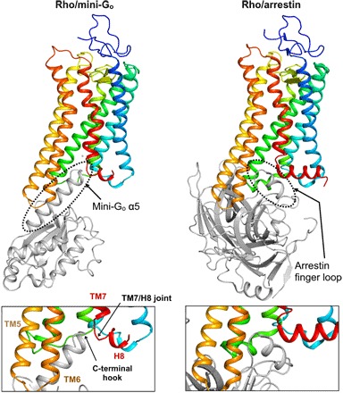
(Top) Comparison of the rhodopsin/mini-Go complex (left) and the arrestin-bound structure (PDB ID: 5W0P; right) shows that rhodopsin exposes an identical site for the recognition of the α5 helix of Gα and the finger loop of arrestin. (Bottom) Detail of the G protein and arrestin binding interfaces viewed parallel to the cell membrane.