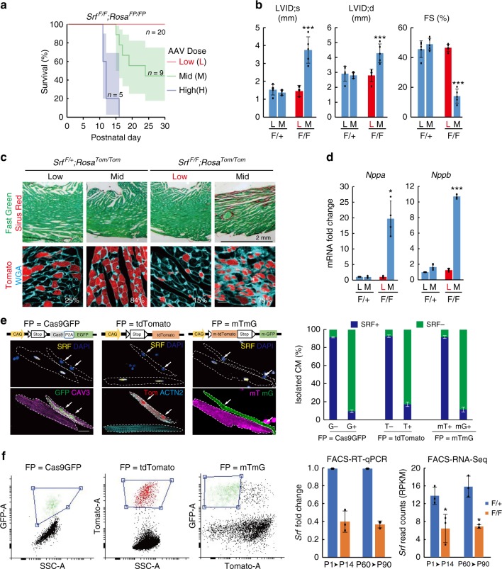
Fig. 1.
Mosaic knockout of Srf circumvents disruption of global heart function. a Survival curve of SrfF/F;RosaFP/FP mice treated with low, mid, and high doses of AAV-Cre at P1. b Effect of AAV-Cre dosage on heart function and chamber size at P30. Left ventricle (LV) function and size were assessed echocardiographically by measuring the LV fractional shortening (FS) and internal diameter at end systole (LVID;s) and diastole (LVID;d). n = 5 per group. c Effect of AAV-Cre doses on myocardial fibrosis. Fibrosis was measured from heart sections stained with sirus red (top) or wheat germ agglutinin (WGA, bottom). The fraction of FP+ cells in heart sections was quantified and labeled in white. d RT-qPCR analysis of cardiac stress marker expression from heart ventricles. n = 4 per group. e Representative images of SRF immunofluorescence in FP+ and FP− SrfF/F;RosaFP/FP CMs that were isolated from the same heart. Arrows point to SRF-FP+ nuclei. Quantification was shown to the right. n = 3 hearts. f Representative FACS plots and gating to sort FP+ CMs (left) and measurement of Srf expression in FACS-sorted FP+ CMs (right) by RT-qPCR and RNA-seq. AAV-Cre was injected at P1 and CMs analyzed at P14, or AAV-Cre was injected at P60 and CMs analyzed at P90. n = 3 hearts. Bar plots show mean ± SD and are overlaid by dot plots of individual data. Two-tailed Student’s t test: *P < 0.05, ***P < 0.001. F/+: SrfF/+. F/F, SrfF/F. Scale bar, 20 µm

