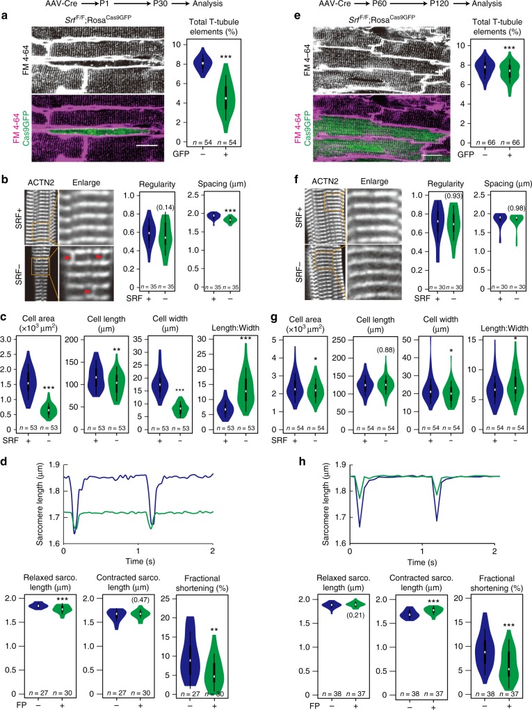
Fig. 2.
Srf plays a profound role in CM maturation but not the maintenance of CM maturity. a–d Mosaic depletion of Srf in neonatal CMs in vivo resulted in dramatic defects in T-tubule organization (a), sarcomere organization (b), maturational hypertrophic growth (c), and contraction (d). e–h Srf ablation in adult CMs had a minor impact on T-tubule organization (e), sarcomere organization (f), maturational hypertrophic growth (g), and contraction (h). a, e Representative in situ images of T-tubules that were labeled by membrane dye FM 4–64 and quantified by the AutoTT software9. Scale bar, 20 μm. b, f Confocal images of ACTN2-immunostained isolated CMs, demonstrating organization of sarcomere z-lines. Red arrowheads point to abnormal ACTN2 staining that extends between z-lines. Regularity was measured by AutoTT and Z-line spacing measured manually. c, g Quantification of the size and morphology of isolated CMs. d, h Measurement of CM contractility. Isolated CMs were paced at 1 Hz. Bright-field images were acquired and then analyzed using SarcOptiM18. In violin plots, white circles show the medians, box limits indicate the 25th and 75th percentiles, whiskers extend 1.5 times the interquartile range from the 25th and 75th percentiles, and polygons represent density estimates of data and extend to extreme values. Two-tailed Student’s t test: *P < 0.05, **P < 0.01, ***P < 0.001. Non-significant P values are labeled within parentheses

