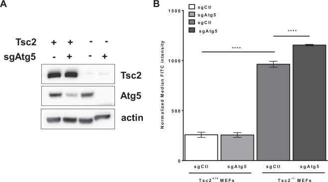
Figure 2.
Macropinocytosis is further increased by autophagy inhibition in Tsc2-deficient cells. (A) Immunoblots confirmed Atg5 knockout in Tsc2+/+ and Tsc2−/− MEFs. The blot was cropped to highlight the relevant bands. The full-length blot is presented in Supplementary Fig. 3. (B) Macropinocytosis was increased in Tsc2−/− sgAtg5 MEFs compared to Tsc2−/− sgCtl MEFs. Median fluorescent intensity of FITC-Dextran was quantified by flow cytometry. Data represented as mean +/− standard deviation of three biological replicates. Statistical significance was assessed using two-way and one-way ANOVAs with Bonferroni correction with ****p < 0.0001.

