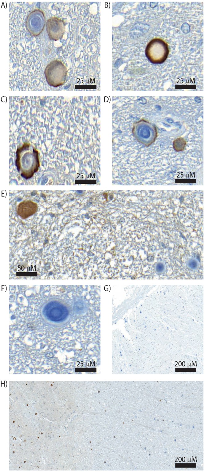
Figure 4.

Detection of misSOD1-positive ring-like structure in the SALS and control individuals Representative images of misSOD1-positive ring-like structures in the extracellular space. (A–D) misSOD1-positive serpiginous structures detected in SALS cases detected in spinal horn grey matter. (E–G) misSOD1-negative CA-like structures detected in SALS cases located in the spinal cord tissue section periphery. (H) Spinal ventral horn grey matter accumulation of misSOD1-positive CA-like structures and, peripheral white matter negative misSOD1 CA-like deposits within the spinal cord.
