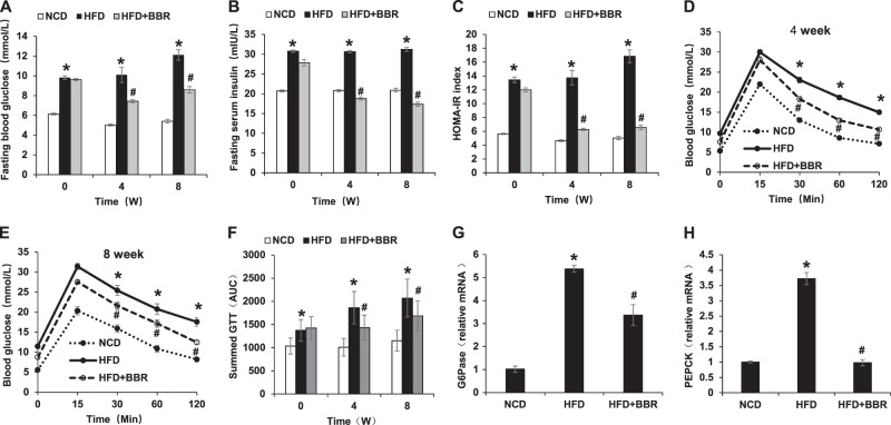
Fig. 2.
Improvement of insulin sensitivity by BBR. a Fasting blood glucose. b Fasting serum insulin. c HOMA-IR. d GTT at 4 weeks of BBR treatment. GTT was performed by intraperitoneal injection of glucose (2 g/kg body weight). e GTT at 8 weeks of BBR treatment. f Area under the curve of GTT assays. g mRNA of G6Pase in liver tissue at 8 weeks of BBR treatment. h mRNA of PEPCK in liver tissue at 8 weeks of BBR treatment. Data are presented as the mean ± SEM (n = 6). *P < 0.05 HFD versus NCD, #P < 0.05 HFD + BBR versus HFD

