Fig. 7.
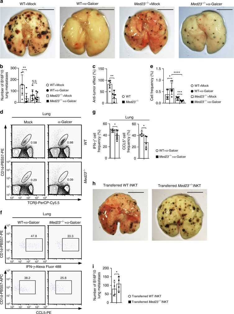
Med23 deficiency impairs the anti-tumor function of iNKT cells. a–c WT and Med23−/− mice were inoculated with 2 × 10⁵ B16F10 cells by i.v. injection. On the same day and again on days 4 and 8, WT and Med23−/− mice received 2 μg of α-GalCer or mock by i.v. injection. Fourteen days after tumor inoculation, the lungs were harvested (a), B16F10 colonies were counted (b) and the anti-tumor effects of α-GalCer were assessed (c) (n = 7). Scale bar: 0.5 cm. The anti-tumor effect (%) = 1-B16F10 colonies (α-GalCer administration)/B16F10 colonies (mock administration). d, e Flow cytometry data (d) and the percentage (e) of iNKT cells in the lungs of WT and Med23−/− mice with post-B16F10 inoculation and α-GalCer or mock administration on days 0 and 4 (WT + Mock, KO + Mock and KO + α-GalCer, n = 8; WT + α-GalCer, n = 7). f, g After in vitro stimulation with PMA and ionomycin for 2 h, flow cytometric analysis (f) was performed, and the percentage (g) of IFN-γ+ and CCL5+ cells among iNKT cells in the lungs of WT and Med23−/− mice with post-B16F10 inoculation and α-GalCer administration on days 0 and 4 was determined (IFN-γ+ cell frequency, n = 7; CCL5+ cell frequency, n = 5). h, i Jα18−/− mice were inoculated with 2 × 10⁵ B16F10 cells by i.v. injection. On the same day and again on days 4 and 8, the Jα18−/− mice received 2 × 10⁵ liver-derived iNKT cells from WT or Med23−/− mice by i.v. injection with 2 μg of α-GalCer. Fourteen days after tumor inoculation, the lungs were harvested (h), and B16F10 colonies were counted (i) (n = 4, paired t-test). Scale bar: 0.5 cm. The data are presented as the mean ± s.d. For all panels: *P < 0.05; **P < 0.01; ***P < 0.001; ****P < 0.0001 by Student’s t-test; N.S. no significance. All data are representative of or combined from at least three independent experiments

