Figure 2.
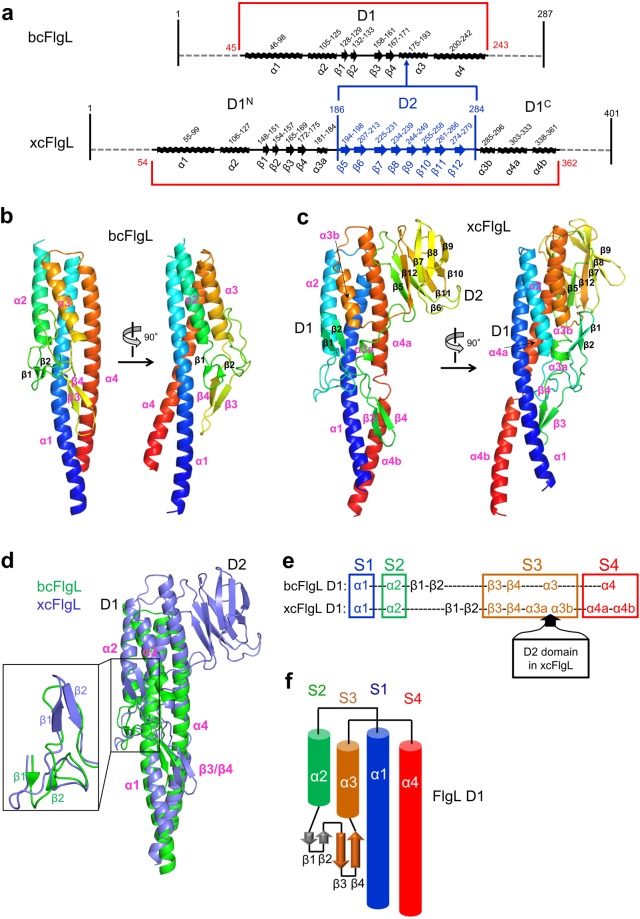
Overall structure of FlgL. (a) Schematic diagram that shows the arrangement of domains and secondary structures in bcFlgL and xcFlgL. (b,c) Structures of bcFlgL (b) and xcFlgLC2 (c). The FlgL structures are shown in rainbow-colored ribbons (N-terminus, blue; C-terminus, red). The secondary structures that bcFlgL and xcFlgL share are highlighted with magenta labels. (d) Overlay of the bcFlgL (green ribbons) and xcFlgL (light blue ribbons) structures. The common secondary structures of bcFlgL and xcFlgL are labeled in magenta. The inset shows that the β1 and β2 strands of bcFlgL are positioned differently from those of xcFlgL. (e) Conserved four-segment (S1–S4) arrangement in the D1 domains of bcFlgL and xcFlgL. The S1, S2, S3, and S4 segments are represented by the secondary structures of α1, α2, β3/β4/α3, and α4, respectively. (f) Topology diagram of the D1 domain in the FlgL structures.

