Figure 4.
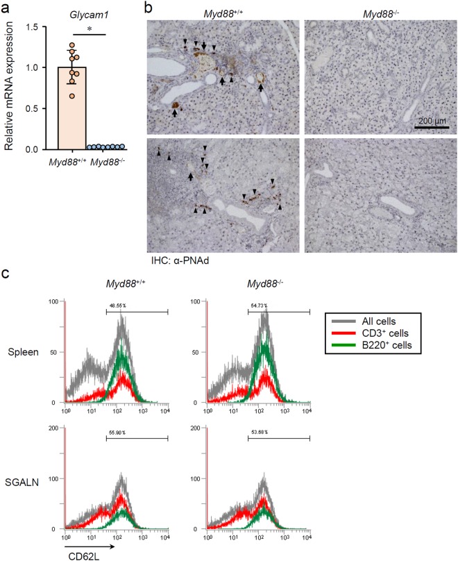
MyD88-dependent HEV formation-associated changes in SGs from NOD mice. (a) qRT-PCR analysis of Glycam1 expression in whole SMG tissues from 12-week-old female Myd88+/+ and Myd88−/− NOD mice (n = 8 per genotype). Expression levels were calculated relative to Hprt expression. Results are expressed as mean ± SD (bar graph). *p < 0.01. (b) Representative areas of anti-PNAd (MECA-79) antibody-stained sections of SGs from Myd88+/+ (left) and Myd88−/− (right) NOD mice. Arrows and arrowheads indicate HEV-like vessels and probable HEV precursor cells, respectively. Original magnification: ×20. (c) Flow cytometric analysis of CD62L in cells prepared from spleen (upper) and SGALNs (lower) from 12-week-old female Myd88+/+ (left) and Myd88−/− (right) NOD mice was performed. Black lines indicate all cells; red lines indicate CD3+ cells, green lines indicate B220+ cells. Results are representative of three independent experiments.

