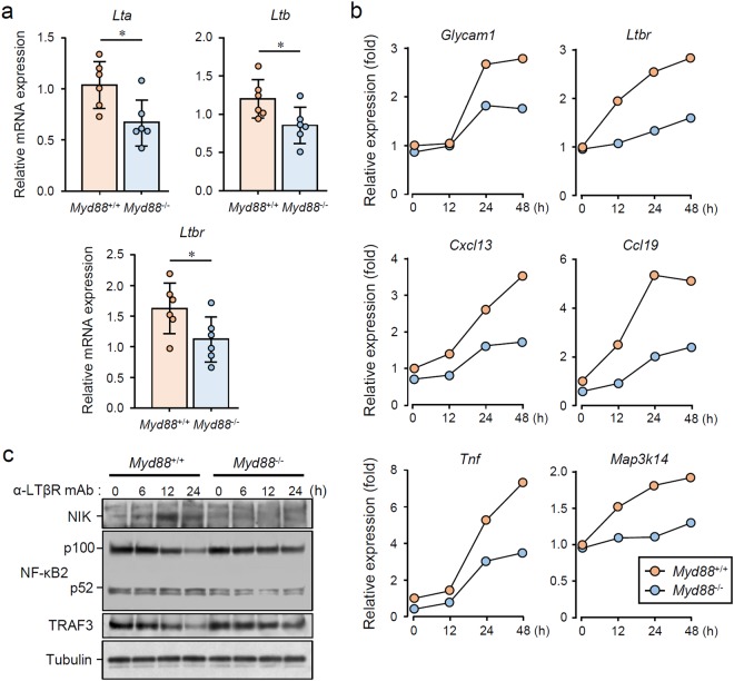
Figure 6.
MyD88-dependent LTβR stimulation-induced expression of TLO-associated genes and signal transduction. (a) qRT-PCR analysis of Lta, Ltb, and Ltbr expression in whole SMGs from 12-week-old female Myd88+/+ and Myd88−/− NOD mice (n = 6 per genotype). Expression levels calculated relative to Hprt expression. Results are expressed as mean ± SD (bar graph). *p < 0.05. (b) Myd88+/+ and Myd88−/− MEFs were stimulated with 2.5 μg/mL LTβR agonistic antibody for indicated periods, followed by total RNA extraction. Relative mRNA expression of Glycam1, Cxcl13, Ccl19, Tnf, Map3k14, and Ltbr was determined by qRT-PCR. Expression levels were calculated relative to Hprt expression. Representative results of three independent experiments are shown. (c) Myd88+/+ and Myd88−/− MEFs were stimulated with 2.5 μg/mL LTβR agonistic antibody for indicated periods, followed by lysis, SDS-PAGE and immunoblotting to assess levels of NIK, NF-κB2 (p100/p52), TRAF3, and Tubulin. Representative results of at least three independent experiments are shown. All the blots were obtained under the same experimental conditions, and the cropped images of the blots are shown. The uncropped images are in Supplementary Fig. 8.

