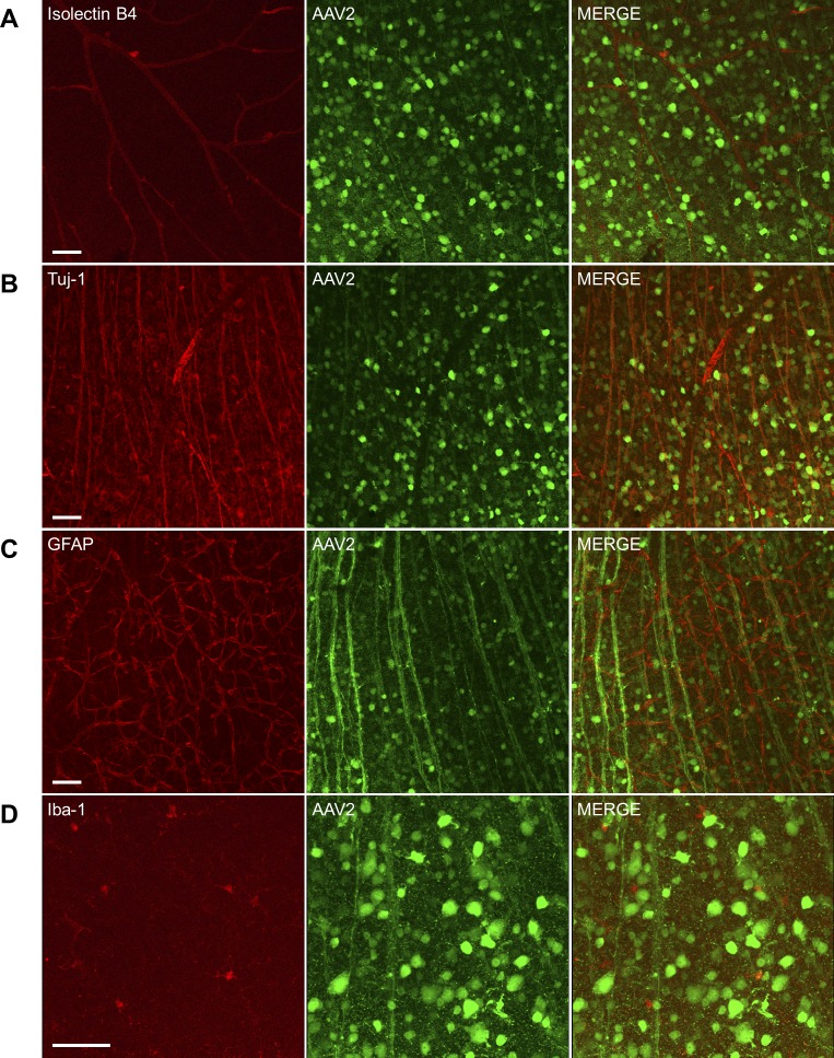
Figure 2.
Transduction characteristics of AAV2-GFP in the retina. Mice received intravitreal injection of AAV2-GFP (1 μL, 1 × 10¹² vector genomes/mL) in both eyes. Eight weeks after injection, retinas were collected and stained with Isolectin B4 (A, red, vessels), anti-Tuj-1 (B, red, RGCs), anti-GFAP (C, red, astrocytes), and anti-Iba-1 (D, red, microglia/monocytes). Confocal images were captured using ×20 (A–C) or ×40 (D) objective. Scale bar: 50 μm.

