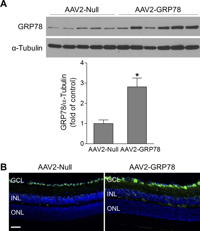
Figure 3.

GRP78 is specifically delivered into RGCs. Retinas or eyeballs were collected at 4 to 5 weeks after intravitreal injection of AAV2-Null or AAV2-GRP78. (A) The expression level of GRP78 was evaluated by Western blotting. α-Tubulin was used as a loading control. Bar graph represents quantification of retinal GRP78 expression. n = 5–6. *P < 0.05 versus AAV2-Null. (B) Representative images of GRP78 immunostaining (green) in retinal frozen sections. DAPI: blue, for nuclei. n = 4. Scale bar: 50 μm.
