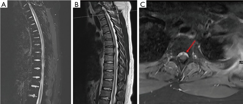
Figure 2.
MRI of the thoracic spine. (A,B) Sagittal cuts of the thoracic spine with T1 post contrast (A) and T2 sequences (B) showing epidural collection along the ventral thecal sac from T1 to T10; (C) axial view at the level of T1–T2 showing epidural collection ventral to the thecal sac with hyperintense material as indicated by the arrow.

