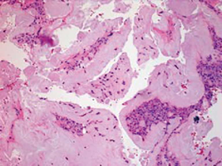
Figure 5.

Histopathology section (HE staining, ×10) of tissue taken from the epidural space at T9–T10 showing fibrous tissue with amorphous crystalline material associated with giant cell reaction consistent with urate gout. However, due to processing of the sample, the crystals were washed out.
