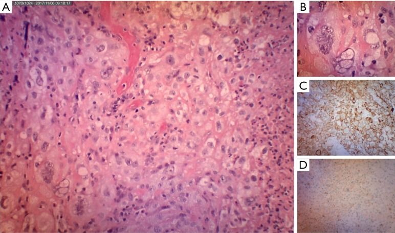
Figure 3.
Histological findings. (A) Sheets and cords of pleomorphic epithelial cells with pleomorphic vesicular nuclei and prominent nucleoli. Adjacent areas of necrosis seen on the right (H&E stain; magnification 10×); (B) giant tumour cells with bizarre nuclei (H&E stain; magnification 40×); these cells are (C) positive for cytokeratin AE 1/3 (magnification 10×) but are (D) negative for thyroid transcription factor 1 (TTF-1) (magnification 10×).

