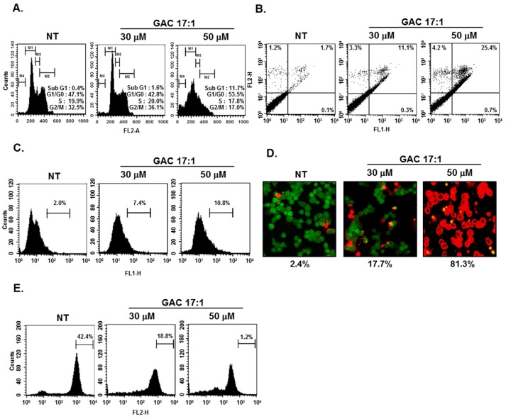
Figure 3.
Apoptotic effects of GAC 17:1 in U266 cells. (A) U266 cells were treated with GAC 17:1 (50 μM) for 24 h. The cells were harvested, washed with a cold PBS buffer and digested with RNase A. Cellular DNA was stained with propidium Iodide and a flow cytometric analysis was done to determine the cell cycle distribution; (B) U266 cells were treated with GAC 17:1 (50 μM) for 24 h and the cells were incubated with Annexin-V/FITC and propidium iodide, then analyzed by a flow cytometer; (C) Apoptosis in U266 cells was detected by TUNEL assay. After treatment, the cells were stained with a TUNEL assay reagent and then analyzed under a flow cytometer; (D) After 24 h of GAC 17:1 (50 μM) treatment, cells were stained with a Live/Dead assay reagent for 30 min and then analyzed under a confocal microscope; (E) U266 cells were treated with GAC 17:1 (50 μM) for 24 h. The cells were washed with a PBS and treated with 5 μM of TMRE (tetramethylrhodamine, ethyl ester) and then analyzed by a flow cytometer to detect MMP activity.

