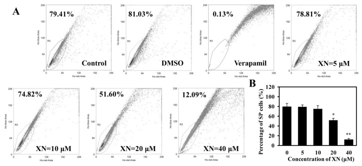
Figure 4.
XN inhibits SP cell percentage in MCF-7/ADR. (A) A representative result of the SP cell percentage is displayed. After treatment with XN (0–40 μM) for 48 h, SP cells were counted by flow cytometry using Hoechst 33342 staining. Verapamil was used to set the SP gate; (B) histograms show the decreasing percentage of SP cells. Data shown are the means ± SD of three experiments for each group. * p < 0.05, ** p < 0.01 versus medium control.

