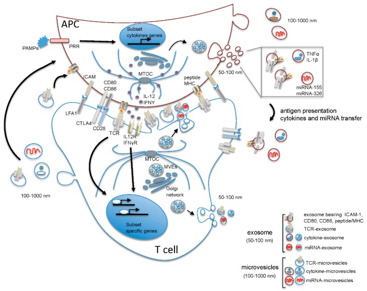
Figure 1.
Suggested model of polarized and non-polarized extracellular vesicle (EV) release during T cell-antigen presenting cells (APC) interaction. Upon contact of the T cell receptor (TCR) with peptide/major histocompatibility complex (MHC) presented by the APC, the segregation of molecules that participate in cell activation occurs at the T cell-APC contact, resulting in the formation of the immune synapse, a highly organized structure characterized by the central accumulation of TCR and peptide/MHC on the T cell and APC side, respectively, and by the formation of a peripheral ring of adhesion molecules (the major being leukocyte function-associated antigen 1 (LFA-1) on T cells and intercellular adhesion molecule 1 (ICAM-1) on APCs), which contribute to consolidating the interaction between T cell and APC leading to the formation of a mature synapse. Intracellularly, the polarization of the microtubule-organizing center (MTOC) to the contact site drives polarized membrane trafficking towards the immunological synapse (IS) and contributes to spatially organize the intracellular signaling and the polarized secretion of soluble mediators into the synaptic cleft. MicroRNA (miRNA)-exosomes and TCR-microvesicles are released from Th cells into the synaptic cleft in a polarized manner, while APC-derived microvesicles and exosomes are released outside the synaptic cleft. Of note, in Th cells, multivesicular endosomes (MVEs) from which exosomes originate are positioned near the contact zone, while in APCs, MVEs do not polarize towards the contact zone, and the release of exosomes and microvesicles occurs in a non-polarized manner. The release of exosomes and microvesicles from T cells outside the synaptic cleft is also shown. Note that the content of EVs has been simplified showing in each vesicle only one of the known components. APC: antigen presenting cell; CD28, CD80, CD86: cluster of differentiation (CD) 28, 80, 86; CTLA4: cytotoxic T-lymphocyte antigen 4; ICAM: intercellular adhesion molecule 1; IL-1β: interleukin 1 beta; IL-12: interleukin 12; IL12R: interleukin 12 receptor; IFNγ: interferon gamma; IFNγR: interferon γ receptor; LFA-1: leukocyte function-associated antigen 1; miRNA: microRNA; MTOC: microtubule-organizing center; MVE: multivesicular endosomes; PAMPs: pathogen-associated molecular pattern molecules; peptide MHC: peptide loaded major histocompatibility complex; PRR: pattern recognition receptor; TCR: T cell receptor; TNFα: tumor necrosis factor alpha.

