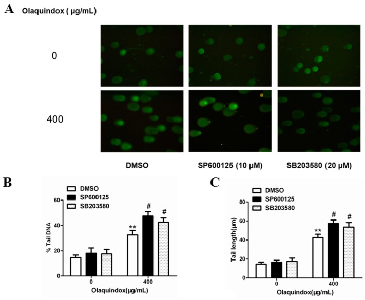
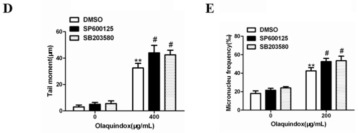
Figure 10.
JNK/p38 pathways played protect role in olaquindox-induced DNA damage. (A) HepG2 cells were treated with olaquindox (400 µg/mL) for 4 h after preincubation with SP600125 or SB203580 for 1 h; (B) % tail DNA; (C) tail length; (D) tail moment; (E) HepG2 cells were treated with olaquindox (400 µg/mL) for 24 h after preincubation with SP600125 or SB203580 for 1 h. Cells were observed under a Leica inverted fluorescence microscope (400×). 1000 binucleated cells were recorded from each experiment and three independent experiments were carried out. All results were presented as mean ± SD. (** p < 0.01, compared with the control group; # p < 0.05, compared to olaquindox group).


