Figure 1.

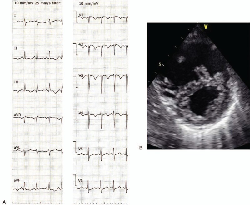
(A) Twelve-lead electrocardiogram on admission showed sinus tachycardia with SIQIII TIII, negative T in V1–3. (B) Short-axis ultrasound cardiogram showed left ventricle compressed by distended right ventricle.

(A) Twelve-lead electrocardiogram on admission showed sinus tachycardia with SIQIII TIII, negative T in V1–3. (B) Short-axis ultrasound cardiogram showed left ventricle compressed by distended right ventricle.