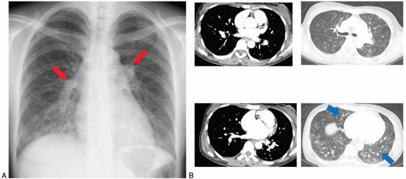
Figure 2.

(A) Chest X-ray on admission showed distended pulmonary arteries (red arrowheads) and bilateral interstitial infiltrate. (B) Chest computed tomography on admission showed no pulmonary embolisms in major pulmonary arteries. Nodular opacities with tree-in-bud pattern are seen (blue arrowheads).
