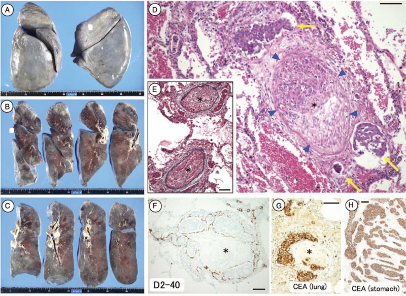
Figure 4.

Macroscopic and microscopic findings at autopsy. No macroscopic nodular lesions were evident in either lung, except for bilateral hilar lymph node metastases (A–C). Distinct fibrocellular stenosis of the pulmonary arterioles was detected histologically (D) (arrowhead). Tumor cells were either detectable or not present within the vessel. Elastica van Gieson staining attributed the fibrocellular stenosis to intimal proliferation (E). Periarterial lymphatics filled with tumor cells (i.e., lymphangiosis carcinomatosa) (D–F, arrow). The lung-infiltrating tumor cells and primary tumor cells in the stomach were both carcinoembryonic antigen-positive (G, H), indicating that the tumor cells in the lung originated from the stomach. Scale bars in D–H: 100 μm. Asterisks in D–G: severe stenotic or occluded pulmonary arterioles.
