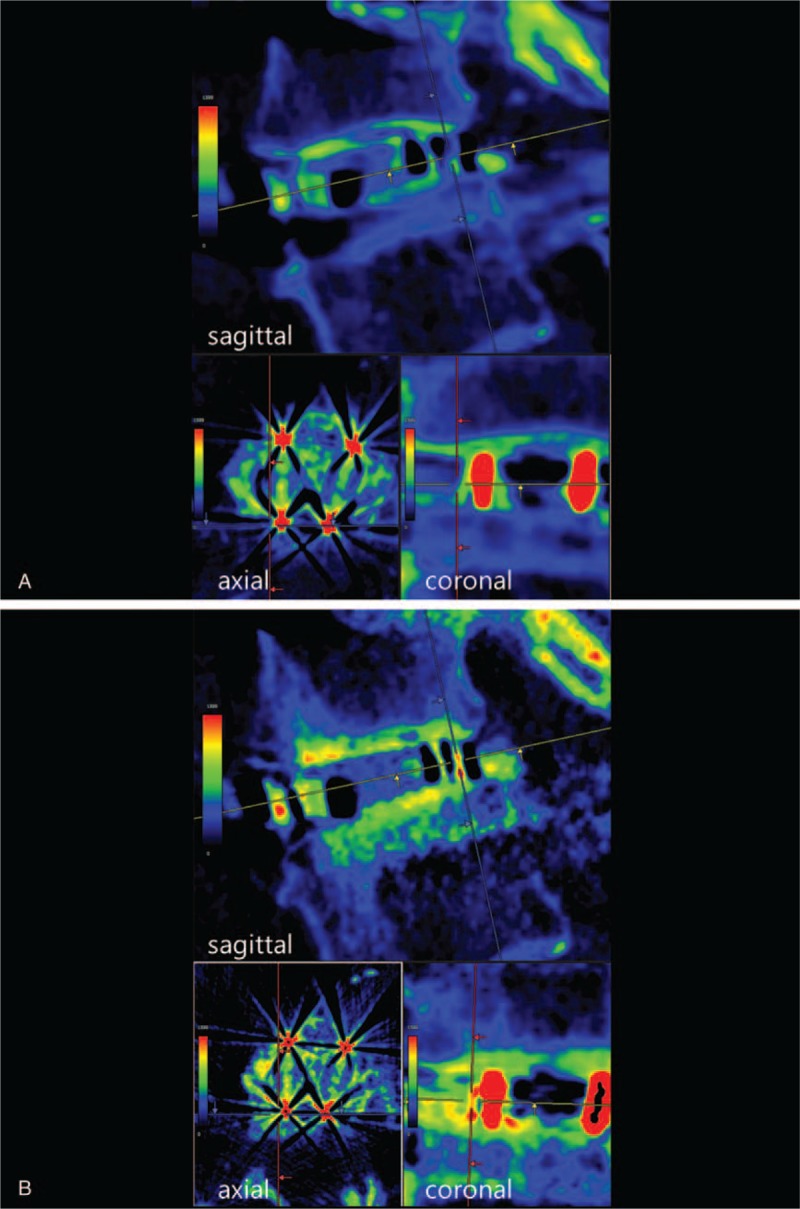
Figure 3.

CT color mapping in a 73-year-old woman who underwent L4–L5 posterior lumbar interbody fusion (A: within 1 week postoperatively, B: 6 months postoperatively). In this case, bone ongrowth was observed on both the upper and lower surfaces of the cage frame in the sagittal plane. CT = computed tomography.
