
Figure 2.
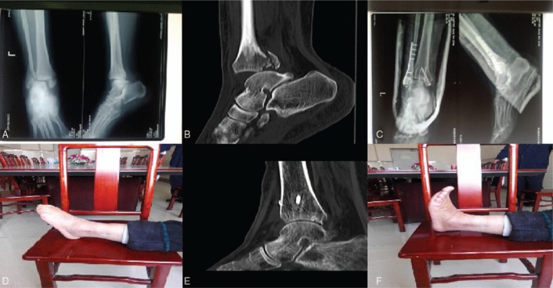
X-ray photograph and CT examination of one patient before and after the ankle joint dislocation. (A) X-ray photograph before the ankle joint dislocation; (B) CT examination before the ankle joint dislocation; (C) X-ray photograph after the ankle joint dislocation; (D) ankle flexion activity one year after the ankle joint dislocation; (E) CT examination 7 months after the ankle joint dislocation; (F) ankle extensor activity one year after the ankle joint dislocation. ∗The patient was female and 58 years old. Sprain led to swelling and pain of right ankle and she was admitted to hospital 2 day after limiting of activities. CT = computed tomography.