Figure 1.

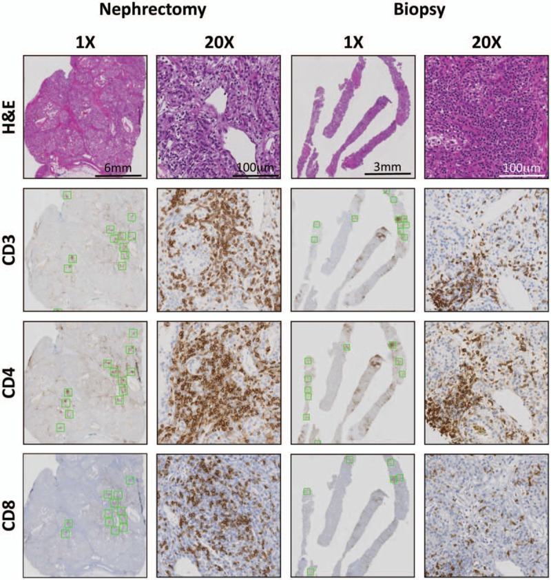
Intratumoral staining and quantification of immune infiltrates in matched nephrectomy and biopsy cases. H&E stain was performed to determine the degree of immune infiltration. Serial slides were stained for T cell markers CD3, CD4, and CD8 to assess the levels of T cell infiltrate in matched nephrectomy and biopsy material from untreated patients. Serial slides (green boxes, shown at ×1) represent areas of highest stain intensity and subsequently quantified for each stain. ×20 images illustrate T cell distribution within an intratumoral immune cell infiltrate in both nephrectomy and biopsy samples.
