FIG 3.
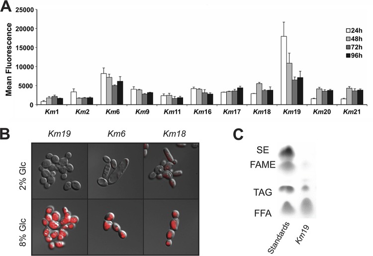
Lipogenesis of K. marxianus strains. (A) Nile red fluorescence flow cytometry of 11 wild-type isolates after 24, 48, 72, and 96 h at 42°C in lipogenesis medium. Experiments were carried out in biological triplicate, with means and standard deviations shown. (B) DIC images superimposed with epifluorescence microscopy of Nile red-stained cells. Little or no fluorescence is seen after 24 h in 2% glucose. After 24 h in 8% glucose at 42°C (Km19 and Km6) and 48 h (Km18), fluorescence is seen encompassing the majority of the cell volume. (C) TLC analysis of Km19 total lipids after 24 h in 8% glucose at 42°C. Lane 1, ladder of standards containing steryl ester (SE), fatty acid methyl ester (FAME), triacylglycerols (TAG), and free fatty acids (FFA). Lane 2, Km19 lipids.

