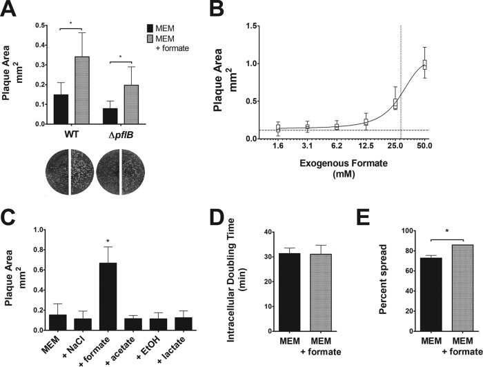
FIG 1.
Formate-induced increase in S. flexneri cell-to-cell spread. (A) Wright-Giemsa stain of Henle-407 monolayers infected with S. flexneri and S. flexneri ΔpflB mutant with or without 20 mM formate. Plaque sizes were measured; an asterisk indicates statistical significance. (B) Plaque size of S. flexneri was measured when various concentrations of formate were added as supplements. The dotted line on the y axis indicates the plaque area when no formate was added as a supplement (0.1 mm2), while the dotted line on the x axis indicates the 50% effective concentration (28.4 mM). (C) Plaque size of S. flexneri was measured with 20 mM NaCl, formate, acetate, ethanol, or lactate; an asterisk indicates a statistically significant difference from MEM. Only formate was found to increase S. flexneri plaque size. (D) S. flexneri intracellular doubling time was measured in Henle-407 cells with or without 20 mM formate. (E) S. flexneri cell-to-cell spread was measured at 3 hpi with or without 20 mM formate; an asterisk indicates statistical significance.

