Fig. 4.
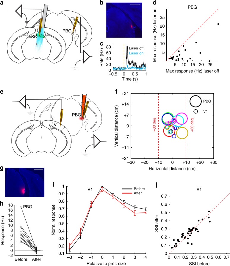
Effect of sSC on V1 is not mediated by PBG. a Laminar recording electrode in the PBG of anesthetized Gad2-cre mouse, expressing channelrhodopsin-2 in the sSC, with a laser-coupled fiber placed above the sSC. b Coronal slice showing trace of DiI (red) from the recording electrode in PBG. DAPI in blue. Scale bar is 0.5 mm. c Example peristimulus time spike histogram in the PBG, without and with optogenetic inhibition of the sSC. d Maximum response to full screen gratings in PBG was reduced by optogenetic inhibition of the sSC (p = 4 × 10⁻⁵, Wilcoxon test, 4 mice, 22 units). e Laminar recording electrodes in V1 and the PBG, before and after injection of fluorescent muscimol in the PBG, in the anesthetized mouse. f Receptive field centers of recorded units in V1 and the PBG for this set of experiments. Colors represent the different experiments. Receptive field sizes are not indicated. g Coronal slice showing fluorescent muscimol in the PBG. DAPI in blue. Scale bar is 0.5 mm. h Muscimol silenced the PBG (p = 0.002, Wilcoxon test; 4 mice, 10 units). i Visual responses in V1 were not changed by PBG silencing (p = 0.57, two-way ANOVA; 4 mice, 43 units). Size is the rank of the stimulus size in a recording, counted from the preferred stimulus in that recording. Error bars represent mean ± s.e.m. j Surround suppression in V1 was not changed by PBG silencing (p = 0.27, paired t-test; 4 mice, 43 units)

