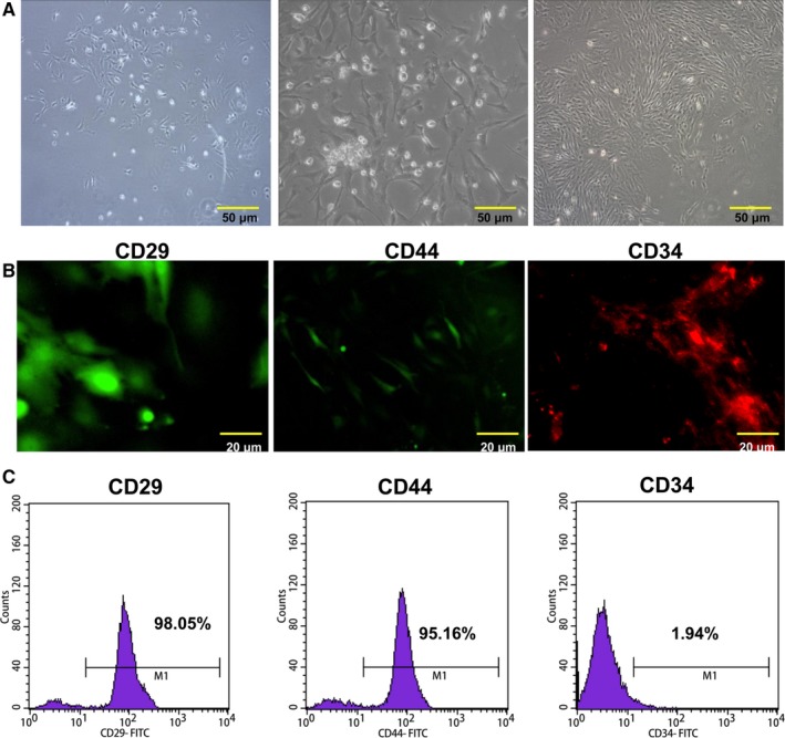
Figure 1.

Characterization of BMSCs. A, The cultured BMSCs. Magnification: ×20, Scale bar = 50 μm. B, CD29, CD44 and CD34 in isolated BMSCs were detected by immunofluorescence assay. Magnification: ×40, Scale bar = 20 μm. C, The isolated BMSCs were characterized by a flow cytometer. BMSCs were positive for CD29 and CD44, but negative for CD34
