
Figure 6.
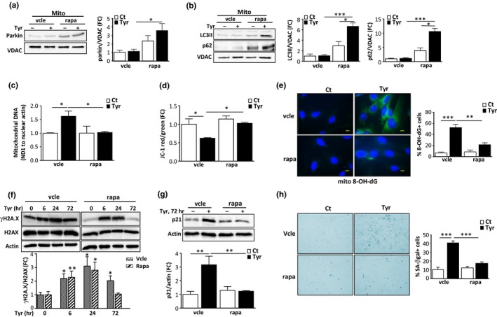
Rapamycin treatment restores mitophagy, preventing the mitochondrial dysfunction, DNA damage response (DDR) and senescence induced by monoamine oxidase‐A (MAO‐A). H9C2 cells were pretreated with vehicle (vcle) or rapamycin (rapa, 100 nM) and stimulated with 500 μM Tyr for 72 hr. (a) Analysis of parkin by immunoblot in mitochondrial extracts; VDAC was used as a loading control for mitochondria (N = 3). (b) LC3 and p62 expression in mitochondrial fractions of cells; VDAC was used as a loading control (N = 3). (c) mtDNA copy number (ND1/actin) by real‐time PCR (N = 3). (d) Quantitative analysis of JC‐1 aggregates (red)/monomer (green) ratios for mitochondrial membrane potential (N = 3). (e) Representative images of mito8‐OH‐dG immunostaining and quantitative analysis of 8‐OH‐dG+ cells as % of total cells (N = 3). (f) Analysis of total and γH2A.X by immunoblot in cells stimulated with Tyr for the indicated times. Actin was used as a loading control (N = 3). (g) Analysis of p21 by immunoblot. Actin was used as a loading control (N = 3). (h) Representative images and quantitative analysis of SA‐β‐gal+ cells as % of total cells after stimulation of the cells with Tyr for 1 week (N = 3). Data are expressed as the mean ± SEM. (*p < 0.05, **p < 0.01, ***p < 0.001)