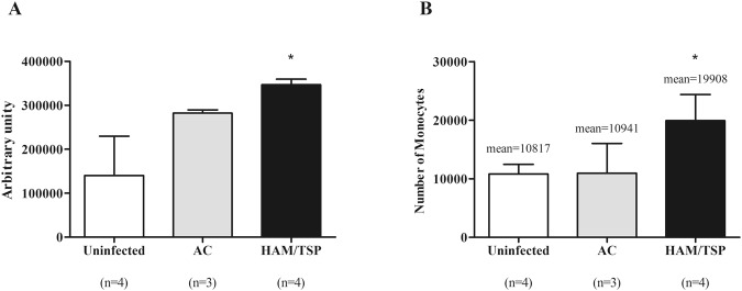
Figure 6.
Monocytes obtained from HAM/TSP patients exhibited higher ability of adhesion and transmigration. (A) Monocytes were labelled with CFSE, washed and resuspended in RPMI medium supplemented with FCS 10% before transfer to BMEC confluent monolayers. Cells were incubated for 3 h at 37 °C with 5% CO2 in humidified atmosphere. Thereafter, quantification of adherent cells was performed by lysing cells with NaOH and measuring the fluorescence intensity. Data are expressed as mean ± SEM of arbitrary units. *Significantly different from the uninfected and AC groups (p < 0.05). The results are representative of four independent experiments. (B) Monocyte transmigration assays were performed in 24-well plates using Millipore Millicell inserts. hBMECs were seeded on the upper side and monolayers reached confluence. Monocytes were incubated for 12 h at 37 °C with 5% CO2 in humidified atmosphere. In sequence, transmigrated monocytes were recovered and counted by flow cytometer using 40,000 fluorescent beads. *Significantly different from the uninfected and AC groups (p < 0.05). Results are representative of five independent experiments, using monocytes from HTLV-1-infected individuals other than those who composed the monocyte pool of the proteomic analysis.

