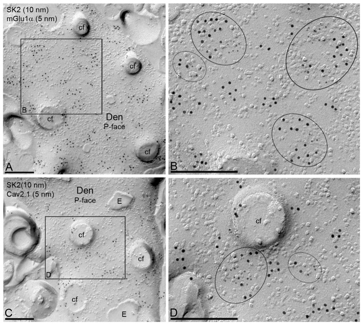
Figure 6.
Co-localization of SK2 channels with CaV2.1 channels and mGlu1α receptor in PC dendritic shafts. Electron micrographs obtained in the molecular layer of the cerebellar cortex showing immunoparticles for SK2, as detected using the SDS-FRL technique. (A,B) Double labeling for SK2 (10 nm) and mGlu1α (5 nm) showing their co-clustering in the P-face of PC dendritic shafts (Den). The black box in panel (A) demarcates the dendritic area shown at higher magnification in panel (B). Clusters of immunoparticles for the two proteins are delineated by black ellipses. (C,D) Double labeling for SK2 (10 nm) and CaV2.1 (5 nm) showing their co-clustering in dendritic spines of PCs. The black box in panel (C) demarcates the dendritic area shown at higher magnification in panel (D). Clusters of immunoparticles for the two proteins are delineated by black ellipses. cf, cross-fracture of dendritic spines; E, E-face. Scale bars: (A–D) 0.2 μm.

