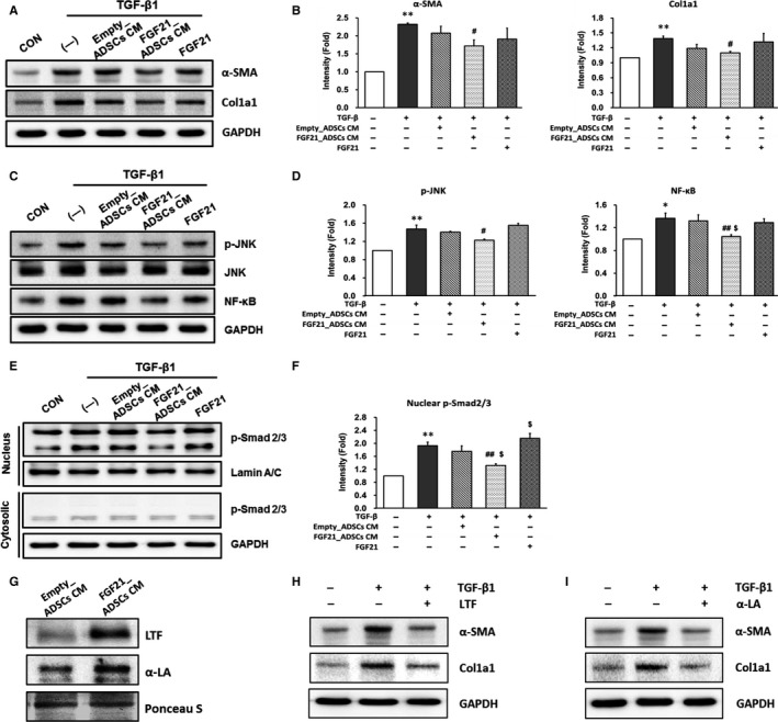
Figure 2.

Effect of FGF21_ADSCs CM on the expression of fibrosis markers in LX‐2 cells and secretome analysis of FGF21_ADSCs culture media. LX‐2 cells were untreated (—) or treated with FGF21_ADSCs CM, Empty_ADSCs CM or 200 pg/mL of FGF21 for 24 h in the presence of TGF‐β1. (A, C, E) Representative western blot and (B, D, F) densitometric analysis for (A, B) α‐SMA and Col1a1, (C, D) p‐JNK, JNK and NF‐κB, (E, F) p‐Smad2/3 and Smad2/3 (n = 3/group). GAPDH or Lamin A/C was used as control for normalization of results. Data are means ± SEM. **P < 0.01, *P < 0.05. vs untreated cells (CON); ## P < 0.01, # P < 0.05. vs TGF‐β1 (—); $$ P < 0.01, $ P < 0.05 vs TGF‐β1 with Empty_ADSCs CM. G, Representative western blot of LTF and α‐LA in Empty_ADSCs CM and FGF21_ADSCs CM. Representative western blot of α‐SMA and Col1a1 (n = 3/group) in TGF‐β treated LX‐2 cells incubated with 100 μg/mL of (H) LTF or (I) α‐LA
