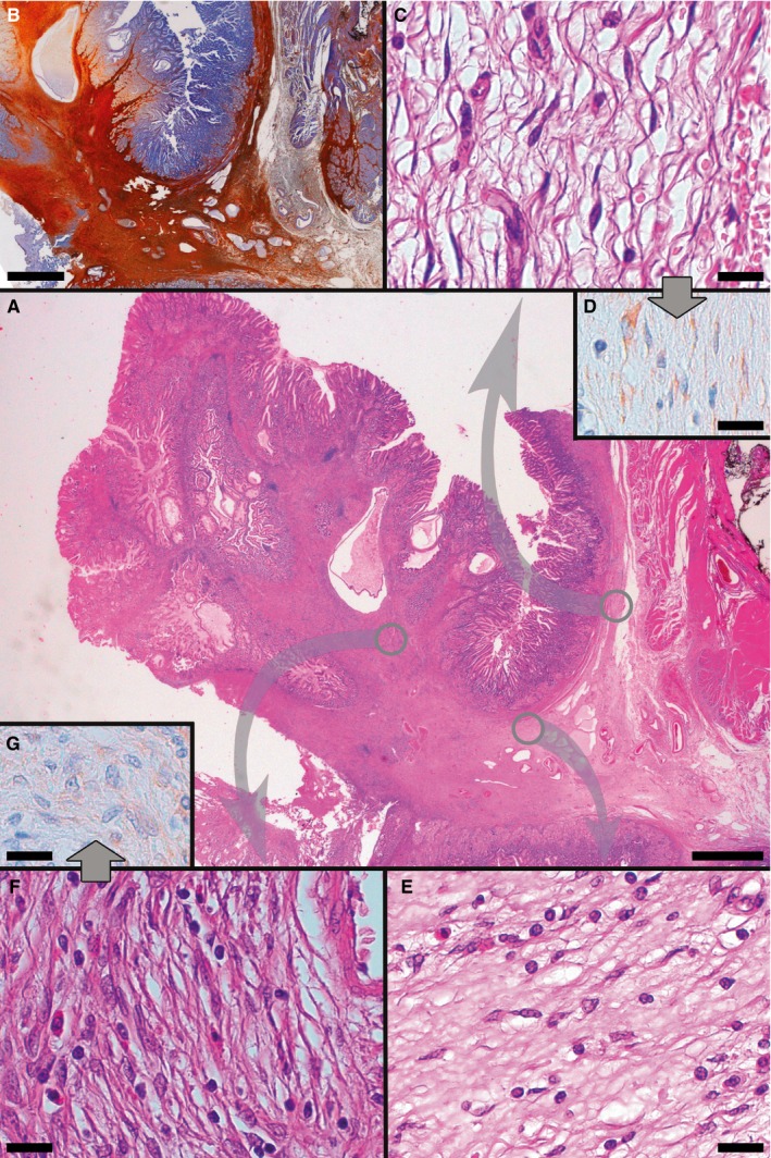
Figure 2.

Representative examples of histological and immunohistochemical transition between TC hyperplasia and IFP found in PDGFRA‐mutant syndrome. At the centre, a panoramic view shows an IFP and its relation with TC hyperplasia (A, H&E). Proceeding in a clockwise direction, both TC hyperplasia and IFP stain positively at CD34 IHC (B). Diverse areas of the specimen are then shown, at higher magnification, in the following panels, which refer to areas encircled in grey in panel A; this is to underline the topographic location of the illustrated microscopic fields to better appreciate the transition between TC hyperplasia and IFP; corresponding areas are connected by arrows. Hyperplastic TCs featuring well‐developed, elongated telopodes, line the outer aspect of submucosa (C, H&E D, PDGFRA). Near the base of IFP, TCs tend to feature shorter cytoplasmic processes, or not to feature them at all (E, H&E). TCs eventually blur into the relatively plump mesenchymal cells characteristic of IFP (note the typical perivascular onion‐skin pattern and the presence of infiltrating eosinophils, absent in TC hyperplasia); PDGFRA positivity is maintained, albeit less intensely than in TC hyperplasia (F, H&E G, PDGFRA). (Scale bars: A, B: 2 mm; C‐G: 25 μm)
