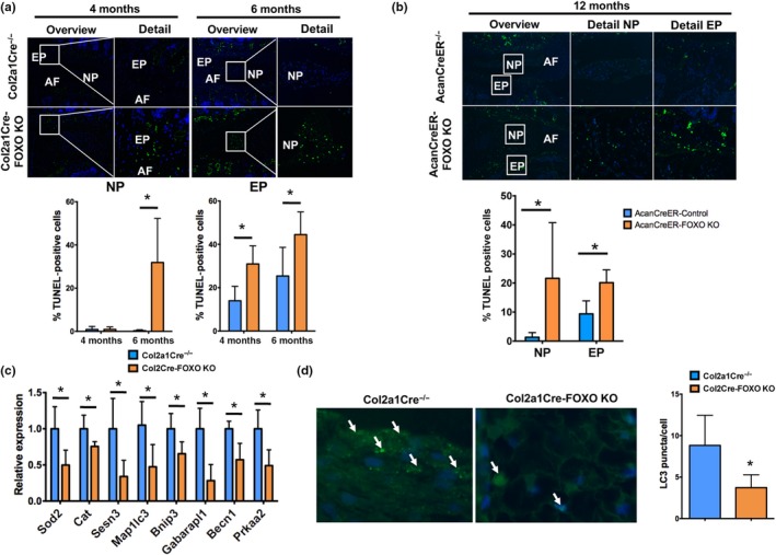
Figure 4.

Reduced nucleus pulposus cell viability and impaired autophagy in FOXO‐deficient intervertebral disks. (a) Terminal deoxynucleotidyl transferase (TdT) dUTP Nick‐End Labeling (TUNEL) staining in lumbar intervertebral disks (IVD) isolated from Col2a1Cre−/− and Col2a1Cre‐FOXO KO mice at 4 and 6 months of age (n = 5 mice per group). Lower panels show quantification of TUNEL‐positive cells in the NP and EP. No positive cells were observed in the AF. NP: nucleus pulposus; AF: annulus fibrosus; EP: endplate. Magnification bar = 100 µm. (b) TUNEL staining in lumbar IVD from AcanCreER−/− and AcanCreER‐FOXO KO mice at 12 months of age (n = 5 mice per group). Lower panels show quantification of TUNEL‐positive cells in the NP and EP. (c) Gene expression analysis of homeostatic genes in NP from Col2a1Cre−/− and Col2a1Cre‐FOXO KO mice at 2 months of age (n = 4 mice per group). (d) Immunofluorescence staining for LC3 in the NP of lumbar IVD isolated from Col2a1Cre−/− and Col2a1Cre‐FOXO KO mice at 4 months of age (n = 5 mice per group) shows a decrease in immunostained cells in FOXO‐deficient NP cells. Right panel shows quantification of LC3 puncta per cell. Values shown are mean ± SD. Statistical comparisons were assessed by an unpaired, two‐tailed t‐test after testing for equal variance using an F‐test. Values are mean ± SD. *p < 0.05
