
Figure 2.
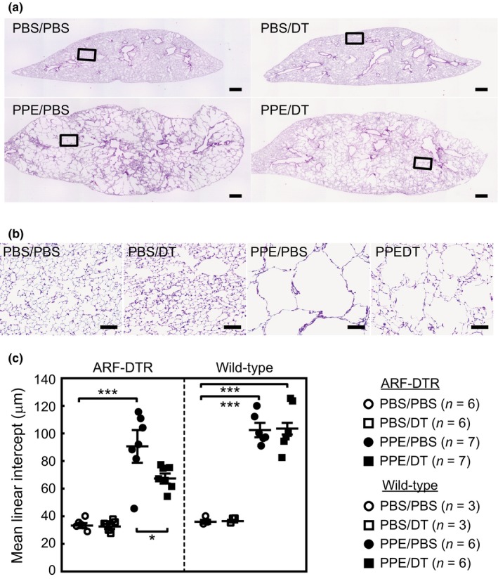
Elimination of p19ARF‐expressing cells diminished elastase‐induced alveolar collapse. (a) Lung tissues of mice treated as shown in Figure 1a were fixed at 25 cmH2O and subjected to hematoxylin and eosin staining. Representative images are shown. Bar; 500 μm. (b) Representative enlarged images of mouse lung sections. The area indicated in panel A was enlarged. Bar; 100 μm. (c) Alveolar mean linear intercepts were measured. At least 300 alveoli were counted in each mouse. Bars represent the mean ± SEM. Data were analyzed by a one‐way ANOVA and Tukey–Kramer post hoc analysis. *p < 0.05 and ***p < 0.001