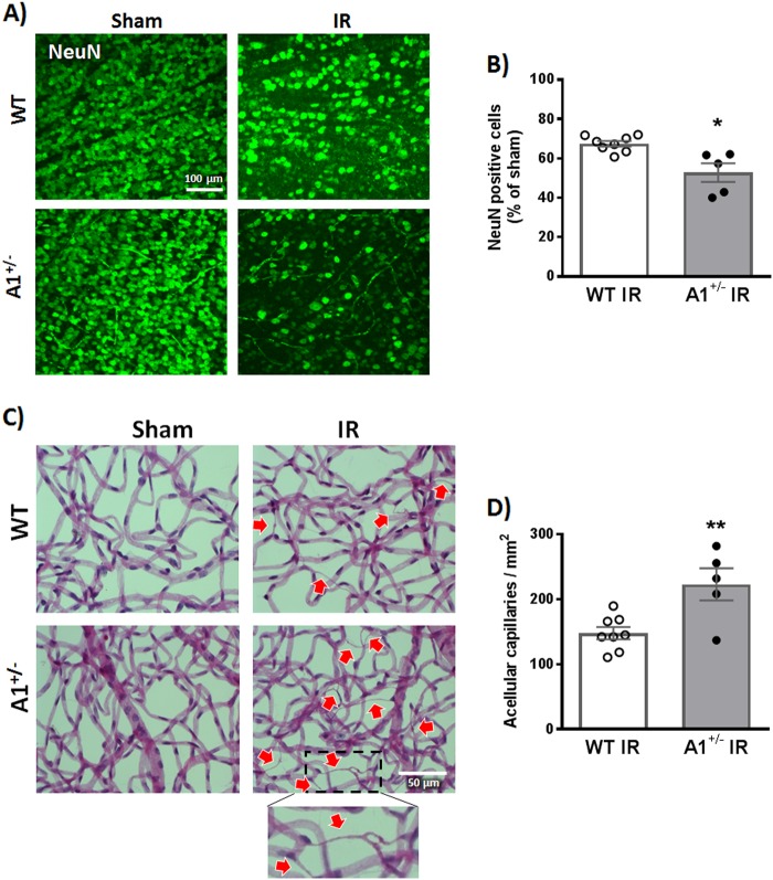
Fig. 1. A1 deletion worsens neuronal and microvascular degeneration after IR injury.
a WT and A1+/− mice were subjected to retinal IR injury and sacrificed at 7 days. Flat-mount NeuN staining showed neuronal cell loss in WT retinas after IR injury compared to shams, which was further aggravated in A1+/− mice. Scale bar = 100 μm. b Quantification of NeuN-positive cells, n = 5 for WT IR and 8 for A1+/− IR, *p < 0.05. c Vascular digests at 14 days showed increased numbers of acellular capillaries (red arrows) in WT IR injured retinas and this microvascular degeneration was further augmented in A1+/− IR injured retinas. Scale bar = 50 μm. d Quantification of acellular capillaries (empty basement membrane sleeves—enlarged in inset), n = 5 for WT IR and 8 for A1+/− IR, **p < 0.01

