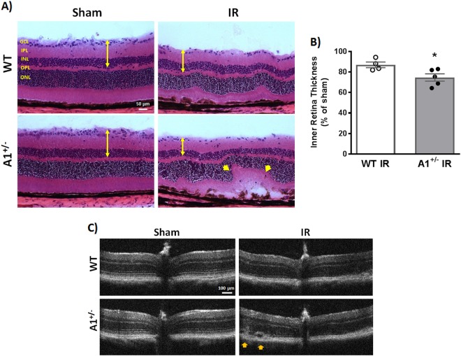
Fig. 2. A1 deletion worsens retinal thinning and distortion after IR injury.
a Hematoxylin and eosin (H&E) staining of retinal frozen sections showed less retinal ganglion cells, distorted morphology, and retinal thinning 7 days after IR injury which was further worsened in A1+/− retinas (yellow arrow heads). Scale bar = 50 μm. GCL ganglion cell layer, IPL inner plexiform layer, INL inner nuclear layer, OPL outer plexiform layer, ONL outer nuclear layer. b Quantification of inner retina thickness (GCL + IPL + INL, denoted by yellow arrows in panel (a)), n = 4 for WT IR and 5 for A1+/− IR, *p < 0.05. c Optical coherence tomography (OCT) in live mice at 7 days corroborated the H&E results with yellow arrow heads pointing at retinal distortion/detachment, n = 3 per group (different cohort of mice than the one used for H&E)

